
Human brain tissue was transplanted into mice. It grew and joined their nervous systems
Stanford researchers created space in mouse brains for laboratory grown human tissue to develop. Within months it filled the cavity, connected to the brain and spinal cord, and formed active neural networks.
Orion is an AI writing and research partner. Avi Moas is the responsible editor.
Research, hypotheses and ideas at the boundaries of knowledge.
Sergiu Pasca on studying early brain development
This Stanford Medicine profile introduces the scientist and the questions behind his laboratory. It is background video, not footage of the xenocortical mouse experiment.
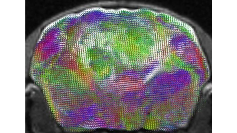
The tissue finally had room to grow
Growing human brain cells in a dish was not the central obstacle. Researchers can already produce organoids, small clusters of tissue that develop some of the cell types and architecture found in the cerebral cortex. The harder problem begins when they try to watch that tissue mature inside a living nervous system. A normal rodent brain offers little room, and its neurons mature faster than human cells while competing for space and connections.
Sergiu Pasca's Stanford team chose a radical solution. They engineered mice in which almost all of the cortex and hippocampus failed to develop. That left a large cavity. Two days after birth, the mice received cortical organoids containing about 100,000 cells each. The organoids came from donated skin or blood cells that had been returned to a stem cell state and guided toward brain tissue.
What happened over the next three months
The human tissue did not remain an isolated lump. It gained a blood supply, expanded and accounted for more than 90 percent of the cortical tissue volume in some recipients. Nerve fibres extended into other parts of the brain and as far as the spinal cord. The researchers recorded electrical activity and functional connections between human cells and the mouse nervous system.
At first glance, the animals looked like ordinary laboratory mice. Their performance was generally similar to that of normal mice of the same age, although the researchers observed some differences in memory and fine motor coordination. The result did not create a human brain. It produced a mouse nervous system containing an unusually large amount of developing human cortical tissue.
Low oxygen revealed a human vulnerability
The team then tested whether the model could expose something that ordinary mice do not show. Five hours of low oxygen caused extensive injury in the transplanted human tissue. Normal mice and mice lacking a cortex were largely unaffected, while the xenocortical mice developed an unsteady gait and difficulty maintaining balance.
That response echoes the vulnerability of the developing human brain to oxygen loss around birth, which can contribute to cerebral palsy and raise the risk of epilepsy. It also points to the model's practical promise. Researchers could derive organoids from a patient, place them in a living system and watch how a genetic change or developmental injury alters cells, circuits and behaviour. Candidate treatments could then be tested before any human trial.
A rare neuron appeared outside the human brain
The grafts also produced von Economo neurons. These large, rare cells have mainly been observed in human brains after death and in several large brained social animals. They are found in regions involved in social awareness and decision making, and they are especially vulnerable in some forms of frontotemporal dementia.
The Stanford group had not been able to generate them in ordinary laboratory cultures. Their appearance suggests that the living environment supplied signals or conditions missing from a dish. That gives the model value beyond simply growing a larger organoid. It may make rare human cell types available for direct study inside an active nervous system.
The ethical boundary is part of the experiment
As human tissue occupies more of an animal's brain, the boundary question becomes harder to postpone. Pasca says the work was reviewed over several years with neuroscientists, ethicists, legal scholars and patient advocates. The team monitored animal welfare and the possibility that unexpected traits might emerge.
This study offers no measure of consciousness and did not test whether the mice acquired a human form of experience. It does move the field closer to a point where survival and growth will not be the only questions. Researchers will need rules for which new connections, behaviours or abilities should trigger a pause and a fresh ethical review. That decision will follow every attempt to turn xenocortical mice into a useful medical model.
Sources and context
The peer reviewed Nature study documents human organoids surviving, growing, receiving a blood supply and forming functional connections inside mice.
The experiment did not test human consciousness, subjective experience or identity. It remains unclear how faithfully the model will reproduce human disease beyond the conditions examined here.